![[Laboratory I -- Introduction to Plant Structure]](AnaD/AnafontN.jpeg)
The plant stele consists of the primary vascular system of the plant axis (stem) and its associated ground tissues (e.g., pith). The stele consists solely of primary tissues differentiated from procambial strands derived from the apical meristem. Secondary vascular tissue (wood consists of secondary xylem) is derived from a vascular cambium.
|
Figure 1.7: Diagram of a plant shoot showing apical meristem, the center of primary growth, a node with leaves and branch bud, and the internode region between nodes. |
Because phloem is rarely preserved on fossils (because the cell walls are not reinforced with lignin, these cells are often crushed or destroyed chemically during preservation), it is the structure of the xylem --particularly primary xylem-- that is of particular interest for paleobotany. Understanding stele types is necessary for interpreting vascular system evolution and for identifying plant axes. The most comprehensive review of stelar morphology in living and fossil plants is that of Beck, Schmid and Rothwell (1982; Botanical Review 48(4):691-817) and a companion paper by Schmid (pp. 817-931) in the same volume. You will note that Rudolf Schmid is a professor in Integrative Biology; he teaches the popular California Plant Life course.
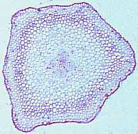
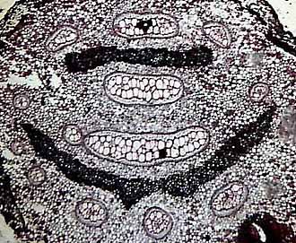
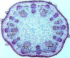
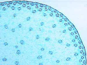
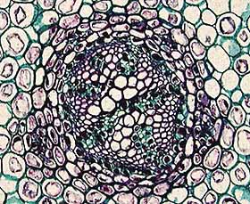
The array of descriptive stele types is overwhelming but don't panic, you don't need to memorize them all. You should become familiar with the protostele (Figure 1.8)(VG 2:1)), a solid interior core of xylem surrounded by a cylinder of phloem, siphonostele (VG 2:3), a central pith (parenchyma) surrounded by a cylinder of vascular tissue, and eustele(VG 2:4)(VG 2:5), separate vascular bundles in the cortex with phloem to the outside of the xylem. This stele is characteristic of dicot angiosperms. You will also encounter dictyosteles, which are complex siphonosteles in which the vascular cylinder is broken up by many leaf gaps. This gives the stem cross section the appearance of concentric, broken rings. Similarly, actinosteles (VG 2:2) are protosteles in which the central vascular strand is lobed, givng is a star-shaped silouette in cross section. There are many other elaborations on these basic steles that will crop up occasionally--be on the lookout!
|
| Figure 1.8: The basic stele types in vascular plants. (A) Protostele, (B) siphonostele, (C-D) eustele. |
One useful character of stelar development is the maturation of the primary xylem. The earliest maturing xylem cells are called protoxylem. These xylem elements are generally small and narrow. Later maturing and larger elements are known as metaxylem. If the protoxylem strands are external to the metaxylem, the stele is exarch (VG 2:6); if protoxylem is internal to the metaxylem, the stele is endarch (VG 2:7); if metaxylem surrounds the protoxylem, the stele is mesarch (VG 2:8).
Combining patterns of xylem maturation with the relative position of phloem and xylem permits a very precise description of the stele in a very few words. For example the sunflower stem (Helianthus) possesses an endarch ectophloic (phloem on the outside) eustele. Do you believe me? Don't look at the slide, check your drawing!
|
| Figure 1.9: Diagrammatic representation of the relationship between stele, leaf trace, and leaf gap in three dimensions.
|
Leaf gaps are features often found in siphonosteles (Figure 1.9). They are discontinuities in the vascular cylinder that occur where leaf traces (the vascular bundles supplying leaves) depart from the stele. If you were to examine serial sections up through the plant axis, leaf gaps would originate and close all along the stele as leaf traces arose at each node.
Leaf gaps occur only in siphonosteles and related types. In protosteles, leaf traces simply diverge from the solid vascular cylinder. The areas between the vascular bundles in a eustele are not leaf gaps. In eusteles, leaf traces arise from individual vascular bundles as if they were tiny protosteles.
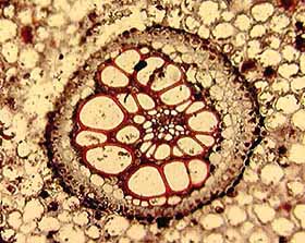
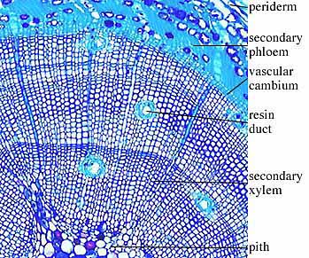
In sections of woody stems, you may note growth rings in the secondary xylem . In seasonal climates, growth varies throughout the year producing annual rings (VG 2:9)(VG 2:10). The diameter of the ring and the size of the cells can tell much about conditions within and between growing seasons. Distinguish primary and secondary tissues in these stems, and note the position of the vascular cambium, although you won't be able to actually see it. Note rays (parenchyma cells revisited) in woody tissue. Diagram (don't draw in cellular detail) the periderm, the protective tissue outside of the wood. Periderm is composed of cork, a secondary tissue derived from activity of the cork cambium.
This is far from an exhaustive review of tracheophyte anatomy and morphology. Undoubtedly many more anatomical terms will come up as our survey of fossil plants continues and as you read the primary literature. Feel free to refer to the several references mentioned here or ask when something seems unclear.
Now, return to the coal ball sections; can you identify plant tissues in the coal balls? What are they? What stele types are present?
![[Previous Page]](../VPLimg/Back.jpeg)
|
![[Title Page]](AnaD/Anabutt.jpeg)
|
![[Glossary]](../VPLimg/Glossbutt.jpeg)
|
![[Next Page]](../VPLimg/Forward.jpeg)
|
![[authors]](../VPLimg/authorbutt.jpeg)
![[copyright]](../VPLimg/copybutt.jpeg)