![[Laboratory I -- Introduction to Plant Structure]](AnaD/AnafontN.jpeg)
The Organism- Building a Plant
|
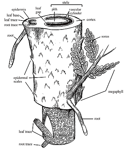
| Figure 1.5: The organ anatomy and general vascular structure of a fern shoot. Note that the megaphyll (= leaf or frond) has abaxial spore-producing structures. Use the three-dimensional diagram to understand how a leaf gap relates to the vascular cylinder of a siphonostele.
|
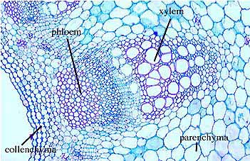
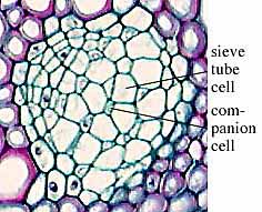
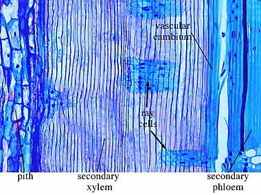
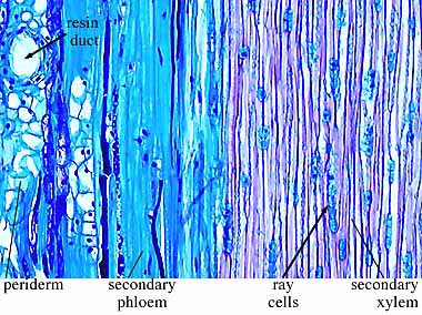
The fern shoot in Figure 1.5 will help you assemble all of the various tissue types into a plant. Examine sectioned stems of modern plants (Helianthus - sunflower (VG 1:5), Pelargonium (VG 1:6), Ranunculus - buttercup (VG 1:7) and Lycopodium (VG 1:9)) to identify parenchyma, xylem, phloem, and other cell types if present. Note that they are arranged together into distinct tissues. If you have never looked at the cellular structure of a plant before, all of the cells may look alike to youÉdon't panic. In modern plant material, the cell types are differentially stained and this will help you distinguish them at first glance. However, don't rely on this crutch because the fossils don't come stained. When you have sorted out which cell type is which, start to notice features of the different cell types that would help you distinguish them in an unstained preparation. For example, phloem cells tend to be a little bit polygonal in contrast to the very round or oval xylem cells. A little time and careful observation (aided by drawing) will train your eye quickly.
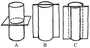
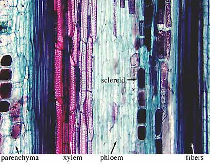
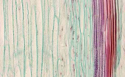
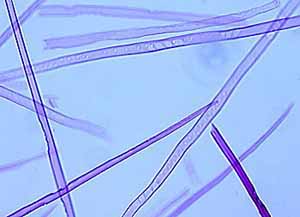
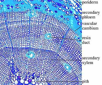
In slides of macerated wood (wood that has been degraded by chemical treatment) you can observe tracheids in three dimensions (VG 1:8). In thin sections of woody stems, note tracheids, fibers, ray cells, and vessel elements (VG 2:9). To find all of these characteristics, observe all three section planes: radial, tangential, and transverse (Figure 1.6) (VG 2:10)(VG 2:11)(VG 2:12). Transverse sections are taken perpendicular to the long axis of the stem (cross-section). Radial sections are taken parallel to the long axis of the stem and cut through the very center of the stem (on radii). Tangential sections are also taken parallel to the long axis of the stem but are cut off center (along a tangent). Each of these views allows you to see the rays in a different orientation. How can you distinguish angiosperm wood from that of conifers and other plants?
|
Figure 1.6: Orientation of sections for the study of wood anatomy. (A) Transverse; (B) longitudinal; (C) tangential.
|
|
Note: Detailed, labeled drawings will be valuable later as you try to recognize these various tissue types in ancient plants. When making drawings, you are trying to compromise between working quickly (so that you can get through all lab material) and providing enough detail to later jog your memory. For example, when drawing a stem cross section, it wouldn't be wise to try to draw every cell. Rather, outline and label the general tissue types (e.g., vascular bundle, ground tissue, cortex), then select one vascular bundle to draw in cellular detail, labeling phloem, xylem, collenchyma, and ground tissue. Artistic merit is not important, utility is. Make sure that your drawings include features important for recognizing the structure or taxon. Also, label your drawings clearly so that anyone (even you when you study for the exam) can interpret them.
|
[Previous Page]
[Title Page]
[Glossary]
[Next Page]
![[authors]](../VPLimg/authorbutt.jpeg) |
![[copyright]](../VPLimg/copybutt.jpeg) |
![[Previous Page]](../VPLimg/Back.jpeg)
![[Title Page]](AnaD/Anabutt.jpeg)
![[Glossary]](../VPLimg/Glossbutt.jpeg)
![[Next Page]](../VPLimg/Forward.jpeg)