Introduction to Placozoa
The Most Simple of All Known Animals
Placozoans are tiny amazing animals.
Very little is known about them because they have never been observed in their natural habitat. No one knows what substrate they live on or what they eat in nature. It is even unknown whether or not they reproduce sexually like most animals. They were discovered in the late 1880's living on the glass walls of an aquarium in a European laboratory. Since then, most of what has been learned about their biology has come from studying cultures of them kept alive in various laboratories around the world. Not surprisingly, given their small size and squishy nature, fossil placozoans have yet to be discovered.
Placozoan from Guam, roughly 400 microns across
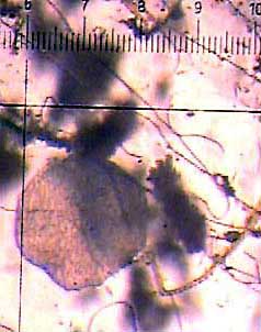
Placozoan Morphology
Placozoans are extremely simple animals. Perhaps not coincidentally, they also have the smallest amount of DNA ever measured for any type of animal. Their bodies are made up of a few thousand cells of just four types. You can compare this to sponges, which have anywhere from 10 to 20 different kinds of cells, to flies, which have roughly 90 different cell types, and to you and other mammals, which have over 200 different types of cells. Placozoans are transparent, flat, round (up to 3 millimeters across), and have two distinct sides. A tissue layer composed of two types of cells, column-shaped cylinder cells with cilia and gland cells without cilia, make up the ventral (or bottom) surface. The upper dorsal surface consists of a layer of just cover cells, which are ciliated and flattened toward the outside of the animal. The image above shows the dorsal surface of a small specimen (just over 4/10ths of a millimeter in diameter) seen from above through a microscope. The dorsal and ventral tissues appear to correspond to ectoderm and endoderm, the outer and inner tissue layers of most animals, but it is not yet known which is endoderm and which is ectoderm. The fourth type of placozoan cells are called fiber cells. These cells are star-shaped and reside in the space between the two tissue layers. The star shape results from thin extensions of the cells which connect to each other in a network. Cellular material such as microtubules and microfilaments traverse the extensions from fiber cell to fiber cell. It is hypothesized that this system of connected cells in important in coordinating the movement of placozoans. Placozoans can move in two ways, by gliding on their cilia and by changing their shape like an amoeba.

Placozoan Feeding and Reproduction
In the laboratory, placozoans have been kept alive by feeding them the flagellated chromist Cryptomonas or the chlorophyte Chlorella. It is unknown what placozoans feed on in nature; they may feed on a number of different organisms. A placozoan feeds with its ventral surface, which produces digestive enzymes. Often, individuals contract part of the ventral surface into a sac where digestion may take place more efficiently. Placozoans can reproduce asexually by either binary fission or, less often, by budding. Some laboratory observations suggest that sexual reproduction may occur. When the population density becomes high, placozoans start to degenerate. Usually a single egg or oocyte develops in the interspace of a degenerating placozoan. Small cells (without flagella) that also form when placozoans degenerate are inferred to be sperm cells. After fertilization, which does not appear to have been documented, cleavage begins. Development has only been observed to the 64 cell stage, at which point the cells cease to separate while the nuclear DNA continues to multiply until the nucleus bursts. Placozoan reproduction and development will probably not be fully understood until they are observed in their natural habitat.

The Phylogenetic Position of Placozoa
Some scientists have inferred that placozoans might be the earliest branch of animals (as shown in box A below) because they are so simple. However, the discovery that placozoan epithelial cells are connected by junctions of extracellular proteins (belt desmosomes), a condition present in all animals other than sponges, suggested that placozoans may have diverged later in the history of animals (box B below). Alternative Hypotheses for the Phylogenetic 
Position of Placozoa
More recently, data from molecular sequences (18S) have indicated that placozoans might have diverged even later in the history of animals (box C above). If this latter view were true, it would imply that placozoans are secondarily simplified from more complex ancestors that had a nervous system. Interestingly enough, placozoans contain cells, which are dispersed around their outer edge, that react with antibodies against a neuropeptide that is present in the nervous system of cnidarians. In any event, the alternative hypotheses for the phylogenetic position of Placozoa within the animals need further testing with additional data.

Placozoan Diversity
Just two species of placozoans have ever been described, Trichoplax adhaerens and Treptoplax reptans. The latter of these has never been seen since its description in 1896, causing some to doubt its existence. The former, however, has been reported from many tropical and subtropical locations around the world, including: the Bermudas, the Caribbean Sea, Eastern Australia, the Great Barrier Reef, Guam, Hawaii, Japan, the Mediterranean Sea, Palau, Papua New Guinea, the Red Sea, Vietnam, and Western Samoa. This prompts the question of whether Tricoplax adhaerens is really a single species. Furthermore, placozoans are so cryptic that their diversity might be much greater than we realize.


Learn more about Trichoplax from Richard L. Howey, whose page "A Weird Wee Beastie", is available at Microscopy-UK.


Sources: