![]() |
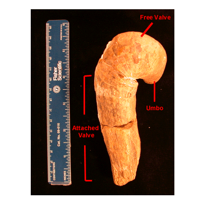
What does a rudist look like? Basic external features of rudists include the umbo and thick, asymmetric right and left valves. The umbo is the rounded protusion found just above the hinge and the hinge is the pivoting point where the two valves meet. There are three main valve plans found in rudists. These plans (from Perkins 1969), are based on relative size of the valves and the level of valve coiling:
|
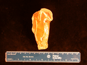
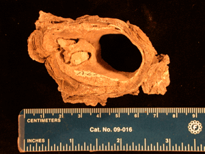
What do they look like on the inside? Important characteristic bivalve features include teeth and sockets which are found in the hinge to prevent shell misplacement, adductor muscles to pull the valves together, and ligamental structures for the movement of the valves. In younger rudist families the ligament function is lost (which will be discussed in more detail later) and there is a question about what replaced this function. Skelton (1976) believes the shell did not open fully and the small opening that existed between the valves was enough for feeding and waste processes. On the other hand, Seilacher (1998) suggests the existence of diductor muscles for opening the valves, but this has yet to be fully proven. Soft tissues are rarely preserved in the fossil record so the study of rudist organs can be a difficult task. |
(Above) Notice the tooth near the center of this valve (from a Masneronia). |
(Above) A longwise section of Coralliachama orcutti. |
(Above) Interior cross section of Monopleura Salazari. |
Evolution of rudist morphology
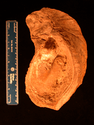
Johnson and Kauffman (1988) summarized important evolutionary changes in rudist morphology. They pointed out that earlier rudists tended to have wide and more coiled bases, while later rudists had thinner bases, more erect forms, and more ornamentation. Sea floor attachment bases became smaller through time while the attached valve went from coiled to more cone-like forms. In later rudist types the free valve became smaller and less coiled.
Important innovation
One important innovation in rudist morphology was the ligamental groove, or invagination of the ligament, which first became distinctive in Caprotinidae (Perkins, 1969). This ligamental groove allowed for more uncoiled shell designs (Steuber 1999). Finally, in Radiolitidae and Hippuritidae, the loss of the functional ligament allowed for an upright growth (Steuber, 1999, Seilacher 1998). For more on these families, see Systematics.
Johnson, C.C., and Kauffman, E.G. (1988), The Morphological and Ecological Evolution of Middle and Upper Cretaceous Reef-Building Rudistids: Palaios, 1988, Reefs Issue, V. 3, p. 194-216.
Perkins, B.F. (1969): Rudist Morphology. - in Moore, R.C. (ed.): Treatise on Invertebrate Paleontology Part N 2 (3) Mollusca 6 Bivalvia - Geol. Soc. Amer. Inc, and Univ. Kansas, p. N751-N763.
Seilacher, A (1998), Rudists as Bivalvian Dinosaurs: Bivalves: An Eon of Evolution--Paleobiological Studies Honoring Norman D. Newell (Edited by Paul A Johnston and James W. Haggart), p. 423-436.
Skelton, P.W. (1976): Functional morphology of the Hippuritidae: Lethaia, v. 9, 83-100.
Skelton, P.W. and Smith, A.B. (2000): A preliminary phylogeny for rudist bivalves: sifting clades from grades: The Evolutionary Biology of the Bivalvia, Geological Society. London, Special Publications, no. 177, p. 97-127.