Bones are the most abundant remains of dinosaurs that we have. And
fortunately, they preserve a lot of information about how the animals
lived. But to get at a lot of this information, you have to get inside
the bones, on the microscopic level.
 In 1994, some generous help from a UCMP donor and field volunteer,
Dr. Jay Grimaldi, enabled the museum to renovate its facility for thin-sectioning
hard tissues. With new tools and machines, we could now examine the
daily growth lines that a snail lays down as it grows and changes its
shell, and we could examine the vascular structures of ancient plant
stems and the insides of fossil seeds. But most of the work that we’ve
done in our lab is in sectioning the bones and teeth of ancient vertebrates.
This work has given us great new insights on how these extinct animals
grew, how they built their skeletons, what they ate, and how active
they were. In 1994, some generous help from a UCMP donor and field volunteer,
Dr. Jay Grimaldi, enabled the museum to renovate its facility for thin-sectioning
hard tissues. With new tools and machines, we could now examine the
daily growth lines that a snail lays down as it grows and changes its
shell, and we could examine the vascular structures of ancient plant
stems and the insides of fossil seeds. But most of the work that we’ve
done in our lab is in sectioning the bones and teeth of ancient vertebrates.
This work has given us great new insights on how these extinct animals
grew, how they built their skeletons, what they ate, and how active
they were.
The science of examining the tissue structures of organisms is called
histology, and it has a long history at UCMP. Charles Camp, J.T. Gregory,
and Frank Peabody were among the scientists who took thin-sections of
the bones of fossil and recent animals to compare their structures.
They started to build this collection in the 1940s, and their slides
are still useful in our work today.
How do we make the thin-sections?
First we choose the tissue we want to sample, be it a shell, a plant
part, or a bone or tooth. At least part of the specimen will be destroyed
by this kind of operation, so usually we make a cast of it first. (After
we’ve sectioned the specimen, we can indicate on the cast where the sections were taken.) Our preparators are so good at this that you can’t even tell what’s been cut after they restore and paint the original specimen. Once we cut out the piece we want to section, we embed it in an epoxy resin—much like superglue—and let it harden. This ensures that the specimen doesn’t crumble and fly apart when it’s sectioned. Then we attach it to a little arm that holds it against the edge of a small, diamond-tipped circular saw blade. This blade cuts slowly but surely through the bone, slicing the specimen like salami in any direction we need. We take these little slices and glue them to glass slides. At this point they’re too thick to let light
|
|
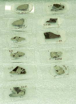
Thin-sections of bone are glued to slides, then ground further until
translucent. (photo by Judy Scotchmoor)
through, so we grind them down on what look like little potter’s wheels
fitted with discs of successively finer sandpaper, until we get the
thinness we need. Then we put them under the microscope.

|