
Living lycopsids are represented by three families: Isoetaceae, Lycopodiaceae, and Selaginellaceae. Isoetaceae and Selaginellaceae both have only a single genus, Isoetes with about 100 species and Selaginella with about 700 species (VG 1:1), respectively. Both of these genera are heterosporous, meaning that each species produces two distinctly different types of spores: microspores and megaspores. The Lycopodiaceae has recently been subdivided into four genera: Huperzia (about 300 species), Phylloglossum (one species), Lycopodiella (about 40 species), and Lycopodium (about 40 species) (VG 1:2). All of these plants are homosporous, meaning they produce only one type of spore. The heterosporous lycopsids, Isoetes and Selaginella, appear to be monophyletic, as is Lycopodiaceae. However, both groups diverged from all other living plants in the very deep past. This phylogenetic pattern probably reflects that the living members are only a small fraction of the group's former diversity. It may also reflect that this diversity had its zenith in the very distant past and has subsequently been extensively cropped by extinction. Think a bit about these issues because they are important for the interpretation of evolutionary patterns from phylogenetic methods, particularly when ancient and/or fossil taxa are involved.
Without exception, the modern lycopsids are low-growing, usually understory or epiphytic plants, herbs; they do not constitute a significant fraction of standing biomass. The greatest diversity of extant lycopsids (as with virtually everything else) occurs in the wet tropics where they live in the understory and are common and species rich as canopy dwellers. Although a relatively inconspicuous component of the modern flora, the lycopsids were arborescent ecosystem dominants in the late Paleozoic.
In general lycopsids, ancient and modern, are characterized by a highly stereotyped anatomy and morphology. Lycopsids have an exarch protostele, actinostele (lobed protostele) or siphonostele (VG 1:3). No living lycopsids show clear secondary growth although some extinct groups did produce a small amount of wood. The interesting genus Isoetes (VG 1:4) produces a suggestion of secondary growth, but it has been so modified as to be difficult to interpret. Branching may be dichotomous or pseudomonopodial. Leaves (microphylls, with only a single vascular strand) and branches are arranged in a phyllotaxic spiral. Many groups (Lycopodiaceae is an exception) have a ligule or small flap of tissue adaxially above each microphyll, or its homologous sporophyll. Sporangia are commonly stalked, kidney-shaped, and are borne adaxially on modified leaves (sporophylls) either clustered in strobili or on fertile portions of the axis (VG 1:5). Stalked, kidney-bean shaped sporangia are the two strongest synapomorphies for the lycopsid clade. Although the living lycopsids look very different and have been evolving as independent lineages for about 250 Ma, they are clearly united to one another and their fossil kin by this character. However, notice the orientation of sporangia on sporophylls. This character allows phylogenetic resolution within the lycopsid lineage.
Lycopsids are first found in the fossil record in the Middle Silurian as part of the "Lower Plant Assemblage" in Australia. The genus Baragwanathia recognized from these beds is clearly a lycopsid based on sporangial form and single-veined leaves, but significantly predates its Northern Hemisphere relatives (VG 1:6). The presence of Baragwanathia (Figure 5.1) and its possible mid-Silurian age have caused significant discomfort and debate among paleobotanists. Considering the early Middle Devonian age of Northern Hemisphere lycopsids, including Baragwanathia, you can understand why. If the mid-Silurian date is correct, how might this fossil record be rationalized in terms of biogeography and evolutionary histories? (Consider what we do and don't know about other mid-Silurian land plants.) Compare specimens of Baragwanathia with the other fossil lycopsids, including Drepanophycus, and also with the living lycopsids. Would you consider Baragwanathia phylogenetically "basal"? How does this alter your interpretation.

|
| Figure 5.1: Reconstruction of Baragwanathia longifolia from the Silurian of Australia. Note sporangial position relative to leaves and how sporangia are concentrated in sections of the stem. Although the age-date of this plant was initially controversial, it has been reconfirmed by several workers and is generally accepted. |
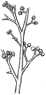
The fossil record of the Northern Hemisphere, where the record is more temporally complete, tells a somewhat different story. Early members of the lycopsid clade include Cooksonia caledonica (Figure 4.4d) from the latest Silurian of Wales, and Renalia hueberi (Figure 5.2C) from the Early Devonian of Quebec. Cooksonia cambrensis is typical of the Cooksonia grade in having naked, dichotomizing axes tipped with reniform sporangia. Renalia is a somewhat larger plant with naked pseudomonopodial axes. Lateral branches dichotomize several times before terminating in pairs of reniform sporangia (VG 1:7). We have excellent Renalia material thanks to paleobotanist Patricia Gensel at the University of North Carolina, who originally described it. Together these taxa, and several others described from the Late Silurian and Early Devonian appear to form a grade of early lycopsids as defined by their typical sporangial shape.

|
Figure 5.2C: Renalia hueberi, whole plant reconstruction. Since Renalia has only been described from compression fossils, nothing is known about its anatomy. |
By the Early Devonian, the kidney-bean-sporangia lineage appears to have split into two distinct groups which become species rich and abundant throughout the Devonian. The zosterophylls differ from Renalia in having many lateral, rather than terminal, sporangia. Most members of this lineage also have the combination of pseudomonopodially-branching main axes or rhizomes, with dichotomous branch tips. Most also have circinate branch tips, which unroll like the fiddle head of a fern.

|
| Figure 5.2: (A) Zosterophyllum deciduum, whole plant reconstruction; (B) ovale protostele of Zosterophyllum. |

|
| Figure 5.3: Vascularization of basal taxa within Lycophyllophytina (sensu Kenrick and Crane, 1997). (A) e.g., Zosterophyllum (naked axis, ovale protostele), (B) e.g., Sawdonia (enations, but no associated vascular traces, (C) e.g., Asteroxylon (enations with vbascular strands extending to base of enation), (D) e.g., Leclercqia (axis bearing vascularized microphylls). |
Much speculation has been offered about the adaptive role of the various epidermal extensions in the zosterophylls. Some speculate that the spines of Sawdonia, and "teeth" of Serrulacaulis and Crenaticaulis protected the photosynthetic axes from predatory arthropods, the remains of which are found in the same beds. Others suggest that protuberances from the epidermis increased photosynthetic surface area on plants that were now growing taller, with thicker stems and more biomass to support. The latter opinion is supported by the presence of stomata on some of the epidermal extensions. At the moment, however, both hypotheses are difficult to test. Furthermore, it is likely that epidermal extensions may have served several functions. What the phylogenetic analysis of Kenrick and Crane (1997) does allow us to say with confidence is that epidermal extensions are derived relative to smooth-axis forms like Zosterophyllum and Renalia.
| Figure 5.4: Sawdonia ornata (A) whole plant reconstruction; (B) sporangia. Protostele can be viewed in Figure 5.3. |

|
![[Title Page]](LycoD/Lycobutt.jpeg)
|
![[Glossary]](../VPLimg/Glossbutt.jpeg)
|
![[Range Chart]](../VPLimg/Rangebutt.jpeg)
|
![[Geologic Time Scale]](../VPLimg/timesbutt.jpeg) |
![[Next Page]](../VPLimg/Forward.jpeg)
|
![[authors]](../VPLimg/authorbutt.jpeg)
![[copyright]](../VPLimg/copybutt.jpeg)